

INTERVENTIONAL RADIOLOGY; FROM DIAGNOSIS TO TREATMENT
Interventional radiology is the department where advanced diagnosis and treatment methods are applied using special equipment (needles, catheters, balloons, stents, drainage catheters, etc.) with vascular and/or percutaneous approaches accompanied by radiological methods.
The following is among the important advantages of the interventional radiology procedures:
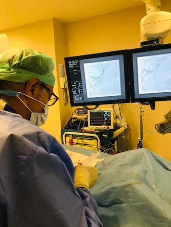
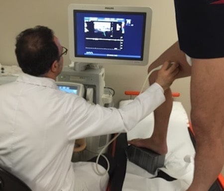
These interventional radiology procedures are applied in combination with ultrasonography, fluoroscopy, Computed Tomography, CT fluoroscopy, digital subtraction angiography (DSA), Magnetic Resonance imaging methods (MR).
Interventional Radiology Procedures
- Oncology– cancer procedures
- Vascular interventional procedures
- Embolization treatments (Vascular Occlusion Therapy)
- Non-vascular interventional procedures directly applied to the skin
Oncology - Cancer Procedures
1. Diagnostic Procedures
A biopsy is the removal of a small piece of tissue or organ to diagnose the disease. Fine needle aspiration biopsy (FNAB) is applied in the form of biopsies tissue or cell/ cytology. The type of biopsy depends on which tissue it is applied to.
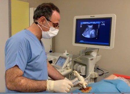
Ultrasonography, computed tomography or magnetic resonance imaging can be used in biopsy as a guiding modality according to localization of the lesion.
2. Port Placement
In order to give chemotherapy to the patient, the catheter is placed in the vein, while the port itself is placed under the skin near the right or left collarbone. This device increases patient comfort.
3. Ablation Treatments in Cancer
Tumor ablation is the application of chemicals or heat (thermal) to destroy the targeted tumor or inflict specific damage.
4. Trans-arterial chemoembolization (TACE)
Occlusion of the artery feeding the tumor by administering chemotherapy (chemo) medication at the same time with high doses of particles through the artery (trans-arterial) feeding the liver is an embolization method applied through chemotherapy.
5. Radioembolization (TARE)
It is the selective application of the radioactive substance (Yttrium-90) - to the point where the tumor is located - into the liver through a catheter placed along the artery by entering through the inguinal artery.
6. New Cancer Treatments
a. Chemo-saturation is applied in liver metastases of melanoma. b. LIOX is applied for the liver cancers
Vascular Interventional Methods
1. Diagnostic Angiography
It is a diagnostic method used for imaging vessels. In the past it was used a routine procedure. Recently It is part of treatment procedures and it is done before treatment.
2. Treatment of arterial stenosis and occlusion
Arteries play a crucial role in transporting oxygenated blood from the heart to all organs and tissues. However, various diseases can lead to narrowing, occlusion, enlargement, or structural abnormalities in these vital vessels. Let’s explore some of these conditions:
Carotid Stenosis and Occlusion:
- The most common cause of carotid stenosis is atherosclerosis, which involves the hardening of arteries due to plaque buildup.
- Stenosis caused by atherosclerotic plaques in the carotid artery can lead to a stroke if left untreated.
- Carotid stenting is recommended in two main scenarios:- When carotid stenosis exceeds 70%, and the patient is asymptomatic or has no brain infarction (dead brain tissue due to insufficient blood supply).
- When carotid stenosis is more than 50%, and the patient experiences symptoms related to brain regions supplied by this artery.
Renal Artery Stenosis:
- Hypertension (high blood pressure) often results from narrowing of the renal arteries.
- Current treatment approaches for renal vascular stenosis involve balloon angioplasty using interventional radiology techniques and stent placement.
Diabetic Foot;
- Diabetic foot complications are frequently associated with atherosclerosis or arterial calcification.
- Diabetes mellitus, characterized by persistently elevated blood sugar levels, can lead to various complaints related to the feet.
Remember that early diagnosis and appropriate management are essential for preventing complications associated with arterial disorders. The treatment approach in diabetic foot patients should cover all stages without delay. These:
- Early (acute) wound intervention,
- Antibiotic treatment,
- Regulation of blood sugar,
- Initiation of blood thinners,
- Elimination of vascular stenosis and obstruction,
- Long-term wound intervention, amputation if gangrene is present,
- Long-term care of the wound,
- Training and implementation of protective measures.
3. Interventional Procedures in Dialysis Patients
Hemodialysis is needed for maintenance of life in patients with impaired renal function. Hemodialysis, or briefly dialysis, is a device used to remove harmful substances accumulated from the body. Dialysis should be performed on a regular basis as often as the patient needs.
Procedures performed on dialysis patients in interventional radiology departments:
- Temporary catheter placement
- Permanent tunneled catheter placement
- Catheter revision and replacement
- Treatment of vein stenosis and occlusion
- Balloon application to fistula stenosis
- Clot removal or clot dissolution in fistula occlusion
4. Deep Vein Thrombosis
The aim of the treatment of sudden leg swelling and the resulting deep vein obstruction and deep vein thrombosis (acute DVT) is to ensure vein patency, prevent clot extension, and reduce the risk of clot throwing into the lungs (pulmonary embolism).
The long-term goal is to prevent the recurrence of clots and to reduce the risk of chronic venous insufficiency and other related diseases (postthrombotic syndrome).
Methods used in treatment;
- Surgical removal of the clot (thrombectomy),
- Thrombolysis, dissolving the clot with medication,
- Catheter-directed clot removal techniques such as catheter aspiration - clot retrieval with a wide-lumen catheter, dispersing the clot with the catheter and breaking it into small particles and eliminating the main vessel obstruction
- Percutaneous mechanical thrombectomy is applied by using high-pressure serum with mechanical devices or by using negative pressure created by rapidly rotating devices to break up the clot and aspirate it at the same time.
5. Varicose Veins Treatment
Varicose veins are a disease of the veins. Veins are the veins that carry the dirty blood to the heart. Unlike arteries, veins have a system of valves. These valves allow the blood to flow to the heart, while preventing the backflow of blood.
In the disease we call varicose veins, blood flows back to the feet due to the deterioration of the valves. The back-flowing blood begins to expand the veins under the skin. These enlarged veins are called varicose veins.
Treatments applied in varicose veins are as follows:
- Laser ablation (burning with laser)
- Radiofrequency ablation (burning off with radiofrequency)
- Microwave ablation (burning off with microwave)
- Foam sclerotherapy (closing the vessel using foam drugs)
- Cyanoacrylate glue ablation (closure using adhesive drugs)
- Treatment of varicose veins with mechanochemical ablation (Clarivein, combination of mechanical and chemical techniques)
6. Aneurysm and Vascular Malformation Treatments
Brain Aneurysm
Brain aneurysm (cerebral aneurysm), known as a bubble in the brain vessels, occurs as a result of weakening of the vessel layers for many reasons such as genetic, traumatic, hemodynamic, or inflammatory.
Although the exact cause is unknown, genetic causes may be the main cause, particularly if aneurysm is detected in several people from the same family. High blood pressure, smoking, trauma are also defined as other risk factors for the development of aneurysm.
Endovascular treatment is the treatment of aneurysm by accessing the artery. The procedure is performed under general anesthesia at the interventional radiology units. Mostly, the intervention site is the inguinal artery, and catheters and microcatheters are made by reaching the aneurysm in the brain with the help of a guide wire. Treatment is applied depending on the preferred method among the catheters. Endovascular Treatment is applied by:
- Simple Coiling,
- Coiling with balloon modeling,
- Coiling with stent modeling,
- Current converter stewing methods.
Peripheral Aneurysms
An aneurysm is the ballooning deformations in the vessels. Vascular ballooning other than the heart and brain is usually defined as peripheral aneurysm. These aneurysms usually develop from arteries in the abdomen. Although it is not seen very often, it is seen in 1-2 out of 100 people. Aneurysms according to the organs in which they develop are as follows:
- Splenic artery (splenic artery),
- Liver artery (hepatic artery),
- Arteries feeding the intestines (gastroduodenal, superior mesenteric artery),
- Liver and spleen common artery (Celiac artery),
- Renal artery aneurysms.
Whereas the treatment of aneurysms was actually surgical in the past, intravascular treatments in interventional radiology, namely endovascular treatments, have come to the fore due to their high success rates and short hospital stays. These treatments are coil embolization, covered stent placement, current-inverting stent placement.
Aortic Aneurysms
The enlargement of the vessel diameters of the aorta - the largest vessel in our body - ruptures and causes life-threatening bleeding. This is why they need to be treated. During the treatment, stent graft applications are performed.
7. Arteriovenous Malformation
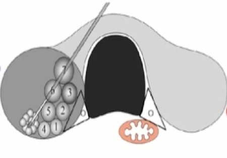
Imperfect development of vessels is called anomaly. There is no normal vascular development and tissue relationship here. Direct association of the artery and the vein without tissue nutrition is called arteriovenous malformation.
While arteriovenous malformation (AVM) often occurs in the brain, it can rarely be seen in other body vessels. Since the capillary structure does not develop in the place where the AVM is located, there is no tissue nutrition and the blood coming from the feeding artery fills the nidus ball and empties into the drainage veins.
Three basic methods can be used in the treatment of AVM. Gamma-knife methods are defined as surgery, vascular occlusion (embolization), and radiotherapy.
AVM embolization is performed by accessing the inguinal artery under general anesthesia at the angiography unit in interventional radiology departments. In embolization, adhesive substances defined as glue or onyx are used.
Embolization Treatments (Vascular Occlusion Therapy)
1. Uterine Fibroid Treatment - Uterine Artery Embolization
Uterine fibroid or myoma uteri is a benign tumor that develops in the uterus. Uterin artery embolization is the recommended treatment method when it is desired to protect the uterus in the treatment of multiple fibroids.
After the catheters are placed in the uterine arteries on both sides, embolization is performed by giving the particles through the catheter. The necessary medical support treatments to relieve the patient’s pain are continued throughout the procedure. Both uterine arteries are occluded.
2. Adenomyosis Embolization
Adenomyosis is a benign disease of uterus and is the uncontrolled growth of the endometrium, the innermost layer of the uterine tissue, through to myometrium.
This causes the uterine tissue to extend and invade the myometrium, the muscle layer. As the disease progresses, it causes deterioration in the myometrium layer.
The procedure is performed with the same technique as for uterine fibroid embolization by accessing through the inguinal artery. After the catheters are placed in the uterine arteries on both sides, embolization is performed by giving the particles through the catheter.
3. Prostate Embolization
Prostate embolization is defined as the interruption of natural blood flow by being blocked by microparticles introduced into the prostate gland through a small catheter.
This procedure is used for the treatment of benign prostatic hyperplasia (BPH-benign prostatic enlargement) to relieve the patient’s complaints of an enlarged prostate gland.
4. Varicocele Embolization
Varicocele is the enlargement of the veins in the scrotum, the bag of skin that protects the testicles in men.
By entering through the inguinal or neck vein, thin tubes called catheters are selectively inserted into the testicular veins. First, venography is applied by administering contrast material through these catheters. Venography shows reflux in the testicular veins, that is, towards the scrotum. After this stage, embolization is applied to the testicular veins by using adhesives called coil, sclerosing substance or glue.
5. Genital Varicose Embolization
This disease, also known as enlargement of the uterine and ovarian veins, genital varicose, pelvic venous insufficiency, female varicocele, pelvic vascular congestion, is the cause of chronic pelvic pain lasting more than 6 months and consists of pelvic venous stasis and pelvic varices.
The point of access to the veins can be the arm, groin or neck vein. Coil embolization of unilateral or bilateral ovarian veins is the most common treatment method in the treatment of ovarian vein leakage - reflux.
6. Hemorrhoidal Artery Embolization
The procedure of occlusion of hemorrhoids, which cause enlargement, by accessing through the arterial tract, is a fairly new, effective, safe treatment that does not require surgery. This treatment is applied by Interventional Radiologists.
7. Hemangioma and Other Vascular Malformation
Embolization of Liver Hemangioma
In its treatment, embolization is performed using percutaneous methods, depending on its content.
Hemangiomas in soft tissue (under the skin, in the muscle) are usually treated by direct skin entry, that is, percutaneously. First, a thin needle is inserted into the lesion and images are taken while a medication called contrast agent is administered. Assessing how much the contrast fills the area in the image and how quickly it empties into the veins, the sclerosant or adhesive medication to be used in embolization is decided accordingly.
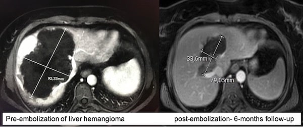
In liver hemangioma, catheter embolization can be done via hepatic artery using particules and chemotherapetic agents. Embolization is a very effective treatment of liver hemangioma.
Liver hemangioma embolization can be done securely in Istanbul and return transportation may be possible within 2 days.
8. Unstoppable Bleeding Embolization
Abnormal bleeding or unstoppable bleeding is an important clinical condition because it carries a life-threatening risk. Common types of bleeding may be listed: nosebleeds, spitting blood, stomach bleeding, intestinal bleeding, bleeding in the urine, vaginal bleeding, bleeding from the previous surgery site.
9. Vascular Access Interventions
Temporary catheter placement (Hemodialysis catheter) Permanent tunneled catheter placement
Non- Vascular Interventional Procedures by Direct Access into the Skin
1. Cyst, Collection Drainage
Pleural Effusion and Acid Drainage
Pleural effusion is the accumulation of fluid in the chest cavity. Percutaneous treatment involves draining fluid accumulations directly from the skin.
Kidney Cyst Drains
Kidney cysts are the most common disease of the kidneys.
2. Abscess Drainage
Collection is the accumulation of fluid in any cavity. When the fluid becomes inflamed, it is called an abscess.
3. Bile Tract Obstructions
It is aimed to remove the obstruction of the bile ducts by entering percutaneously. This blockage can occur for benign causes such as stones and biliary sludge, or for malignant causes such as cancer of the biliary tract, gallbladder, duodenum, and stomach. In cases where surgical methods cannot be applied to remove the stones, percutaneous methods are applied. After percutaneous entry into the biliary tract, the stone is pushed into the intestine using a balloon occlusion catheter. Transhepatic balloon application to the level of the ampulla also contributes to the treatment.
Bile Duct Drainage - Percutaneous transhepatic cholangiography, it is the visualization of the bile ducts by giving contrast material with a needle after entering the bile ducts. Occlusion is assessed after PTK. It is the reduction of bilirubin values by delivering bile to the intestine with the help of a multihole catheter or taking it out of the liver in biliary tract obstruction.
Internal biliary drainage: Reaching the intestine by passing the stenosis or obstruction in the bile ducts with a catheter and transferring the bile to the intestine.
Biliary stent: A plastic or metallic tube (cage) inserted endoscopically or directly through the skin as an interventional radiology approach to relieve stenosis or obstruction in the biliary tract. Metallic stents have a longer efficacy time. While plastic stents can be removed from the biliary tract endoscopically, metallic stents are permanent.
4. Urinary Tract Obstructions
Stone disease is one of the diseases that cause obstruction in the urinary tract. Other causes include benign and malignant tumors. Tumors may occur at the exit level of the kidney, along the ureters, or originate from the bladder. Causes originating from the bladder are initially unilateral, but over time, depending on the growth of the tumor, obstruction in the urinary tract that causes enlargement of the collecting system in both kidneys may occur.
Urinary tract obstruction treatment can be done in 2 ways:
Percutaneous Nephrostomy; When urine cannot reach the bladder due to obstruction in the urinary tract, enlargement of the urinary tract develops in the kidney, this is called hydronephrosis. A catheter is inserted through the percutaneous route, directly through the skin, entering the renal collecting system. The catheter is attached to the bag outside, allowing urine to accumulate in the bag. General anesthesia and hospitalization are not required in these day-to-day procedures.
Double J catheter or ureteral stent placement: Hydronephrosis develops due to stenosis and obstruction in the ureters. If hydronephrosis is not treated in the early stages, deterioration in kidney functions occurs. The stenosis and obstruction in the ureters are treated with the bladder entrance from the bottom up or by the percutaneous way directly into the kidney by the antegrade way, that is, the top-down way.
5. Non-Cancer Ablation Techniques
Thyroid Ablation
In this technique, under the guidance of ultrasound, a thin needle is inserted through the skin into the nodule in the thyroid and the nodule is destroyed by the heat generated by the laser and radiofrequency. Radiofrequency ablation is a reliable alternative to surgery in benign thyroid nodules.
Breast Fibroadenoma Ablation
Cryoablation therapy in breast fibroadenoma ablation is a method approved by the FDA in 2002. In cryoablation, the mass is reached with a fine needle and cooled down to -80 degrees. Due to freezing, the cells in the fibroadenoma die, and the mass shrinks within months and becomes impalpable. It is a safe alternative to surgery.
Ablation of Benign Bone Tumors (Osteoid Osteoma, Osteoblastoma)
It typically develops from the outer part of the long bones, namely the cortex. The tumor is usually close to the joint, that is, diaphyseal or metadiaphyseal. It is a benign tumor surrounded by bone reaction (osseous sclerosis) and (reactive) bone edema, with or without white spot (central mineralization) with (osteolytic) nidus formation, which is typically seen as black on computed tomography, usually smaller than 15 mm. It is safely treated with ablation.
Soft Tissue Tumors Ablation
Ablation can be applied in cases of recurrence of some tumors that develop in the soft tissue. Diseases such as desmoid tumor and endometrioma located under the skin can be treated with cryoablation